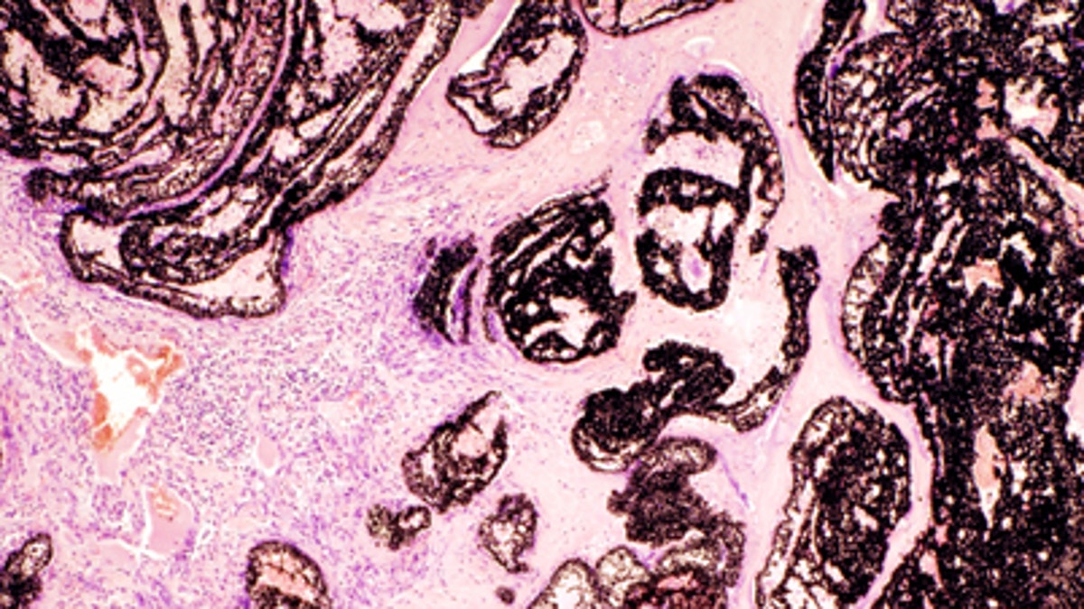
(SpringerImages.com/Bielefeld Hospital)
Cancer researchers are teaming up with astronomers using computerized algorithms designed for viewing distant galaxies to spot biomarkers that can indicate the aggressiveness of a tumor, Medical News Today reported.
The method pathologists currently use for finding out the aggressiveness of a tumor involves looking through a microscope at a stained tumor sample to identify different proteins and cell changes. A computerized system would speed up the process.
In a study published in the British Journal of Cancer, teams from the Cancer Research U.K. Cambridge Institute, and both the Department of Oncology and the Institute of Astronomy at the University of Cambridge in the U.K. looked at more than 2,000 breast cancer tumors to determine the levels of three proteins known to be biomarkers for aggressive cancer.
"We've exploited the natural overlap between the techniques astronomers use to analyze deep sky images from the largest telescopes and the need to pinpoint subtle differences in the staining of tumor samples down the microscope," lead author, Dr. Raza Ali, a pathology fellow from the Cancer Research U.K. Cambridge Institute, said in a statement.
Researchers analyzed each tumor sample both manually with a pathologist using a microscope and automatically with a computer using adapted algorithms. When they compared the results, they found the computer algorithms came to the same conclusion as the pathologists up to 96 percent of the time.
"The results have been even better than we'd hoped; with our new automated approach performing with accuracy comparable to the time-consuming task of scoring images manually, after only relatively minor adjustments to the formula," said Ali.
The researchers now plan to do a large-scale, international study, using tumor samples from more than 20,000 breast cancer patients.
"Modern techniques are giving us some of the first insights into the key genes and proteins important in predicting the success or failure of different cancer treatments,” said senior author Carlos Caldas, a professor at the Cancer Research U.K. Cambridge Institute. “But before these can be applied in the clinic, their usefulness needs to be verified in hundreds or sometimes thousands of tumor samples."
New methods are already helping researchers analyze up to 4,000 images a day, Caldas added.


