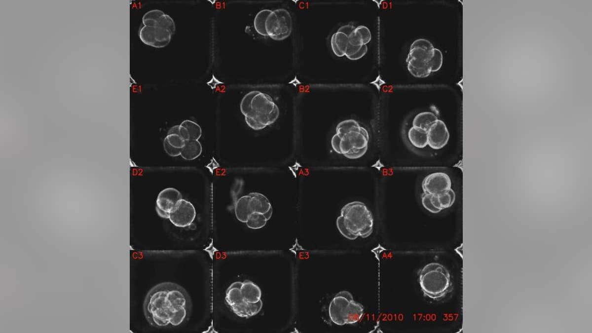
Time-lapse images of human embryos in the first two days of development. (Kevin E. Loewke)
Amazing time-lapse videos of embryos in the very earliest stages of development could help fertility doctors prevent miscarriage, new research suggests.
By watching the timing of the cells' development, doctors could determine which cells are genetically healthy, and which have abnormal numbers of chromosomes, finds the study published today (Dec. 4) in the journal Nature Communications.
Chromosomes are coiled packets of DNA. Humans have 23 pairs of chromosomes, but genetic accidents can alter that number, a condition called aneuploidy. Some aneuploidies cause disorders such as Down syndrome, which occurs when there are three chromosomes on what should be the 21st pair. Other aneuploidies are incompatible with life, causing early miscarriage or later stillbirth.
Extra or missing chromosomes are shockingly common, affecting up to 75 percent of all embryos, studies find. This may be why as many as 50 to 75 percent of pregnancies are so-called "chemical pregnancies," meaning that an embryo spontaneously aborts right after implantation in the uterus. Many women with chemical pregnancies may not even realize they were ever pregnant.
There's little to be done about these early miscarriages in typical pregnancies. For in vitro fertilization (IVF), however, it's important to choose embryos with the best chance of life to prevent miscarrying.
Renne Reijo Pera, a Stanford University stem cell researcher, and her colleagues have already found that abnormal embryos show strange behaviors in the first four days of development. For example, the length of time it takes an abnormal embryo to complete its very first division from one cell body to two differs from the time it takes for a normal embryo to do the same.
The researchers wanted to know whether they could use these odd behaviors to reliably distinguish a healthy embryo from a doomed one. They took 75 human embryos that had been frozen at the single-cell phase and cultured them in Petri dishes for two days, taking a microscopic snapshot of each embryo every five minutes. [See Video of the Developing Embryos]
These snapshots were then strung together into time-lapse movies, which the researchers analyzed for the timing of various cell-division phases.
Of the 75 original cells, 53 survived four days, which represents the zygote stage of embryonic development. Of those, 45 were usable for genetic analysis. About 75 percent, or 34 of the 45 cells surviving to the zygote stage, had the wrong number of chromosomes.
The abnormal cells showed more variations in their cell-division cycles than normal cells, the researchers found. While normal cells all developed at similar paces, abnormal cells lagged behind or sped ahead in the divisions of the first, second and third cells.
Combining data about the abnormal timing with other signs that something has gone wrong (such as fragmented DNA and asymmetrical cell sizes within a developing embryo) could reliably show which cells have the right number of chromosomes and which don't, the researchers report.
Distinguishing between aneuploid embryos and those with 23 pairs of chromosomes "may contribute to improvements in IVF outcomes by potentially reducing the inadvertent transfer of embryos that would most likely result in embryonic lethality and spontaneous miscarriage," the researchers wrote.
Follow Stephanie Pappas on Twitter @sipappas or LiveScience @livescience. We're also on Facebook & Google+.
Copyright 2012 LiveScience, a TechMediaNetwork company. All rights reserved. This material may not be published, broadcast, rewritten or redistributed.
