High-tech tool to boost fertility
Fertility doctors at the Cleveland Clinic and 15 centers around the country are using a new camera to revolutionize in-vitro fertilization
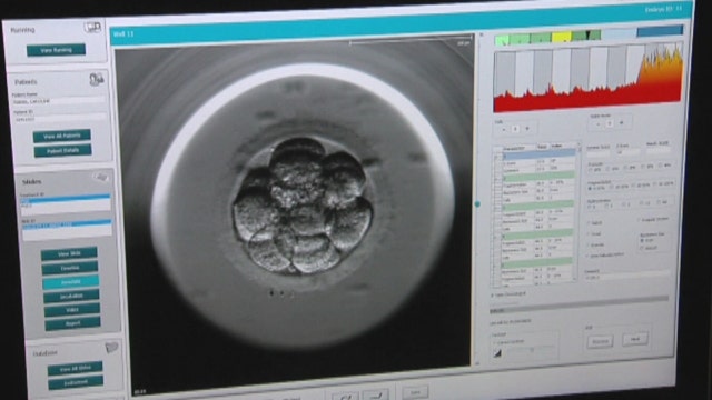
A newly developed technology called the embryoscope is changing the playing field for fertility doctors across the country.
Traditionally, during in-vitro fertilization, doctors will check embryos once a day to determine whether or not they are viable. However, this process is tricky because it must be done in a quick and efficient manner – embryos must be kept at 37 degrees and in an environment with the proper concentration of gases in order to survive.
However, with the embryoscope, a picture can be taken of the embryos every few minutes without disturbing them in their incubators.
Dr. Nina Desai, of the Cleveland Clinic in Ohio, says having the embryoscope in her facility has been helpful.
“Embryos are extremely dynamic for a moment to moment, hour to hour, they will change,” Desai said. “And those changes can reflect a lot about the competence of an embryo to implant on the uterine wall.”
More than 100 patients at the Cleveland Clinic have been able to start families thanks to this high-tech tool and it is now being used at 14 other medical centers across the country.
"I think for patients it must be an incredible experience to be able to hold a baby in their arms and to then be able to go back and look at this video of their embryos from the very first minutes, first hours of the embryo growing before it even implanted in the uterus," Desai said.
The embryoscope has the power to potentially revolutionize in-vitro fertilization and research is currently being done to determine what role this technology is playing in increasing fertility rates.

