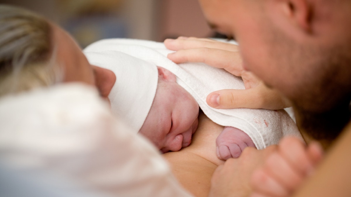
Newborn baby girl right after delivery, shallow focus
As of today, there are no definitive tests to measure a child’s risk for developing autism. Since early intervention and therapy is key for at-risk children, such a test could be critical for managing the early development of a child.
Now, researchers at Yale School of Medicine and the MIND Institute at the University of California, Davis say they have found a safe and effective way to measure a newborn’s risk for developing an autism spectrum disorder (ASD) – by looking at his or her placenta.
In a new study published in the online issue of Biological Psychiatry, senior author Dr. Harvey Kliman and his colleagues examined abnormal placental folds and cell growths called trophoblast inclusions, which acted as effective biomarkers for predicting which children were at risk for developing ASD.
“There are no methods at birth to diagnose this at all. Period,” Kliman, a research scientist in the department of obstetrics, gynecology and reproductive sciences at Yale, told FoxNews.com. “The only advanced notice that a family might have a child with autism spectrum disorder is that they have a previous child (with autism) – which is sort of unfair, because it’s a high price to pay.”
One out of every 50 children is diagnosed with autism in the United States each year, according to the latest report from the Centers for Disease Control and Prevention (CDC). However, most children are diagnosed with ASD at the age of 3 or 4 – long after their first year of life, when early intervention can be the most effective.
A serendipitous discovery
Kliman said he wasn’t originally looking to develop an autism test, and he stumbled upon the placenta connection “totally by accident.” When he first started working at Yale, his main job was to examine the tissue of lost pregnancies – to better determine why the baby didn’t make it. A main component of the tissues he analyzed were the chorionic villi found in the placenta.
Chorionic villi are tiny, finger-like structures that help transport blood between the mother and the developing fetus. The villi are covered by a layer of cells called trophoblasts, which – much like skin – help to protect the structure’s insides. Normally, the trophoblast layer is smooth, but if the cells grow abnormally, the layer can fold on itself forming what are called trophoblast inclusions.
“What the textbooks said was if you have trophoblast inclusions, it means you have a triploid pregnancy – which is a single egg that is fertilized by two sperm,” Kliman said. “If you have two sperm fertilizing at the exact same time, you wind up having 69 chromosomes,” or three sets of chromosomes.
“But to confirm, we’d do a test to see if they had three sets of chromosomes, but they would often have two sets of chromosomes,” Kliman added, “and that’s not what the textbooks said.”
Doing more research into trophoblast inclusions, he found they weren’t simply an indication of a triploid pregnancy, but they were also signs of genetic abnormalities – including conditions like Down syndrome and abnormal heart conditions.
The discovery ultimately got him thinking: If these placental folds are a sign of genetic abnormalities, is there a difference in the frequency of trophoblastic inclusion among those at risk for autism?
Beyond the fold
To test this theory, Kliman and his colleagues examined 217 placentas – 117 placentas from infants born to at-risk families and 100 control placentas. Children of at-risk families – those who have had a previous child with autism – are nine times more likely to be diagnosed with autism than the rest of the population.
Of the at-risk placentas, some had as many as 15 trophoblast inclusions, a great deal more than the control placentas, which never had more than two inclusions. Ultimately, the team found a placenta with just four trophoblast folds equates to a 96.7 percent probability that the infant is at risk of developing autism.
As for why autism is linked to these abnormal placentas, the scientists do not know for sure, but they do plan to study the underlying mechanisms of this connection moving forward.
“We believe that there is a generalized biological problem in the way the cells are interacting in the bodies of these people,” Kliman said of their theories. “The placenta is part of the baby and is simply a reflection of this difference. But it’s something to do probably with things that regulate cell growth and how cells divide.”
Kliman noted this test – which they have aptly named the PlacentASD test – does not diagnose children with autism, and it cannot be done prenatally, meaning it cannot be used as a screening exam during pregnancy. But he said the test can be a very useful tool for parents, giving them a better understanding of how their child is developing during those first few years of life.
“If you’re positive in this test, what I would say to the family, is your child has the same risk of having autism as a family who already has a child with autism,” Kliman said. “Because that is a significantly increased risk over the normal population, it would be worthwhile to have increased surveillance and vigilance about your child. Also, you might want to contact people who do early intervention.”
Early intervention techniques often include more attentive parenting methods – such as making sure the child responds to stimuli normally and closely monitoring that the child is hitting major developmental milestones. Kliman added that no child has ever been harmed by these kinds of techniques, so when you ultimately weigh the test’s pros and cons, there are really no downsides to having it done.
“This is a very simple test,” Kliman said. “No one needs the placenta after delivery. We’re not asking anyone to give a difficult biopsy or go through any pain, so from that point of view it’s very easy.”


