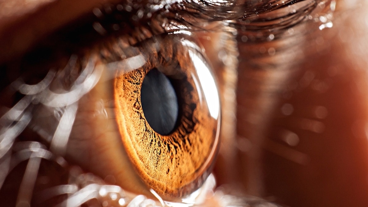
(air009/Shutterstock)
In a medical first, surgeons have used a robot to operate inside the human eye , greatly improving the accuracy of a delicate surgery to remove fine membrane growth on the retina . Such growth distorts vision and, if left unchecked, can lead to blindness in the affected eye.
Currently, doctors perform this common eye surgery without robots. But given the delicate nature of the retina and the narrowness of the opening in which to operate, even highly skilled surgeons can cut too deeply and cause small amounts of hemorrhaging and scarring, potentially leading to other forms of visual impairment, according to the researchers who tested out the new robotic surgery in a small trial. The pulsing of blood through the surgeon's hands is enough to affect the accuracy of the cut, the researchers said.
In the trial, at a hospital in the United Kingdom, surgeons performed the membrane-removal surgery on 12 patients; six of those patients underwent the traditional procedure, and six underwent the new robotic technique. Those patients in the robot group experienced significantly fewer hemorrhages and less damage to the retina , the findings showed.
The technique is "a vision of eye surgery in the future," Dr. Robert E. MacLaren, a professor of ophthalmology at the University of Oxford in the United Kingdom, who led the study team and performed some of the surgeries, said in a statement. MacLaren presented the results today (May 8) at the annual meeting of the Association for Research in Vision and Ophthalmology (ARVO), happening this week in Baltimore.
"These are the early stages of a new, powerful technology," said MacLaren's colleague Dr. Marc de Smet, an ophthalmologist in the Netherlands who helped design the robot. "We have demonstrated safety in a delicate operation. The system can provide high precision [at] 10 microns in all three primary [directions], which is about 10 times" more precise than what a surgeon can do, de Smet said. (The three primary directions are up/down, left/right, and towards the head/towards the feet.)
Membrane growth on the retina results in a condition called epiretinal membrane, a common cause of visual impairment . The retina is the thin layer at the back of the eye that converts light waves into nerve impulses that the brain then interprets as images.
An epiretinal membrane can form because of eye trauma or conditions such as diabetes, but more commonly it is associated with natural changes in the vitreous, the gel-like substance that fills the eye and helps it maintain a round shape. As people age, the vitreous slowly shrinks and pulls away from the retinal surface, sometimes tearing it.
The membrane is essentially a scar on the retina. It can act like a film, obscuring clear vision, or it can distort the shape of the retina. The membrane can form over the macula , a region near the center of the retina that sharply focuses images, a crucial process for reading or seeing fine detail. When membranes form here, a person's central vision becomes blurred and distorted, in a condition called a macular pucker.
Removing the membrane can improve vision , MacLaren said, but the surgery is very intricate. The membrane is only about 10 microns thick, or about a tenth the width of a human hair, and it needs to be dissected from the retina without damaging the retina … all while the eye of the anesthetized patient is jiggling with each heartbeat, MacLaren said.
Faced with the need for such precision, de Smet and his Dutch-based group developed a robotic system over the course of about 10 years. Robot-assisted surgery is now commonplace, particularly for the removal of cancerous tumors and diseased tissues, as in the case of hysterectomies and prostatectomies. But it has never been tried on the human eye, given the finer precision needed, the researchers said.
De Smet's group had a working model of the robotic system in 2011, devised by de Smet and Maarten Steinbuch, an engineering professor at the University of Eindhoven in the Netherlands. They demonstrated the system's utility in 2015 on pigs, which have similar size eyes as humans.
MacLaren's team first used the system on a human, a 70-year-old priest from Oxford, England, in September 2016. Upon the success of that surgery, MacLaren's team conducted a study on 11 more patients in a randomized clinical trial, hoping to measure the robotic system's accuracy compared to the human hand.
The robot acts like a mechanical hand with seven independent motors that can make movements as precise as 1 micron. The robot operates inside the eye through a single hole less than 1 millimeter in diameter and goes in and out of the eye through this same hole during various steps of the procedure. But the surgeon is in control, using a joystick and touch screen to maneuver the robot hand while monitoring movements through the operating microscope, MacLaren explained.
During the trial, two patients who underwent the robotic surgery developed micro-hemorrhages, which means a little bit of bleeding, and one experienced a "retinal touch," which means there was an increased risk of retinal tear and detachment. In the traditional surgery group, five patients experienced micro-hemorrhages, and two had retinal touches.
MacLaren said the precision offered by the robotic system may enable new surgical procedures that surgeons have dreamed about but figured were too difficult to accomplish. For example, MacLaren said he hopes to next use the robotic system to place a fine needle under the retina and inject fluid through it, which could aid in retinal gene therapy , a promising new treatment for blindness.
"The robotic technology is very exciting, and the ability to operate under the retina safely will represent a huge advance in developing genetic and stem cell treatments for retinal disease," MacLaren told Live Science.
The surgical system was developed by Preceyes BV, a Dutch medical robotics firm established at the University of Eindhoven by de Smet and others.


